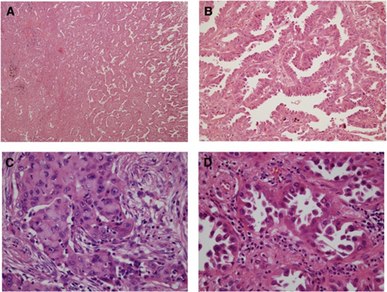
Figure 2.
Representative images of RET-rearranged adenocarcinoma of the lung. (A and B) Many RET-rearranged adenocarcinomas displayed a papillary growth pattern (A: low magnification, and B: high magnification). (C) Solid signet-ring cell pattern was observed in a minority of RET-rearranged adenocarcinoma (original magnification × 200). (D) Some tumour cells displayed homogeneously eosinophilic-to-pale inclusions in the nuclei (original magnification × 200).

