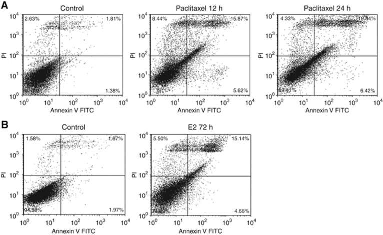
Figure 2.
Differential apoptotic effects of E2 and paclitaxel. MCF7:5C cells were treated with control or (A) paclitaxel (1 μM) for 12 and 24 h or (B) E2 (1 nM) for 72 h, and then stained with annexin V-FITC and PI and analysed by flow cytometry. Viable cells (left lower quadrant) are annexin V-FITC− and PI−, early apoptotic cells (right lower quadrant) are annexin V-FITC+ and PI−, dead cells (left upper quadrant) are PI+ and late apoptotic cells (right upper quadrant) are annexin V-FITC+ and PI+. Increased staining for apoptosis is observed maximally in the right upper quadrant.

