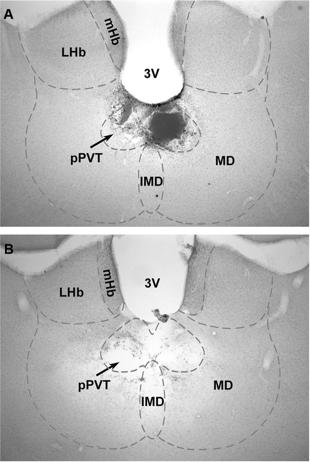
Figure 1.

Examples of lesions of the pPVT. The location of the lesions as verified from histological sections of the brain were used to group the data into pPVT lesion (complete lesions limited to the pPVT); (A) and midline lesion (complete lesion of the pPVT and surrounding area including the mediodorsal and intermediodorsal nuclei); (B) groups. 3V, third ventricle; IMD, intermediodorsal nucleus; LHb, lateral habenular; mHb, medial habenular; MD, mediodorsal nucleus; pPVT, posterior paraventricular nucleus of the thalamus.
