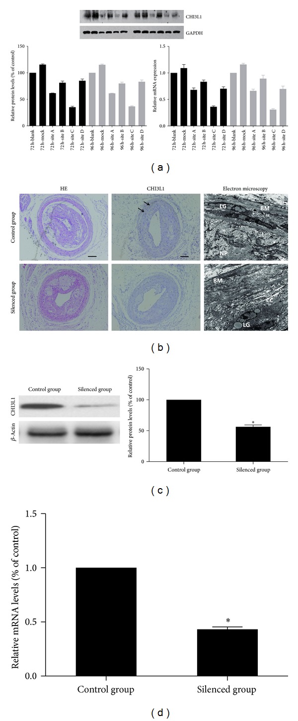
Figure 2.

(a) Target site screening for CHI3L1 by western blot analysis and real-time RT-PCR in RAW264.7 cells. The RAW264.7 cell line was transfected with lentivirus expressing different CHI3L1 siRNAs, and gene silencing analysis showed that site C lentivirus was the most effective vector in blocking CHI3L1 expression. (b) The immunohistochemical staining of CHI3L1 and electron microscopy in control group and silenced group. In the control group CHI3L1 expression (arrow) could be demonstrated according to the immunohistochemical staining. However, little CHI3L1 was expressed in silenced group. For electron microscopy, in control group most of the endothelial cells denudated and there were a large number of lipid granules (LG) under the basement membrane (BM) in the vessel wall. The atherosclerotic plaques were occupied with necrotic particles (NP), calcification crystals (CC), and cellular debrises. However, in silenced group the number of lipid granules was relatively decreased. (scale bars = 100 μm) (c) Western blot analysis and quantification of CHI3L1 protein expression in control group and silenced group. The levels of CHI3L1 protein expression were higher in control group than in silenced group. (d) Real-time RT-PCR quantification of CHI3L1 mRNA expression in control group and silenced group. *P < 0.05 versus control group.
