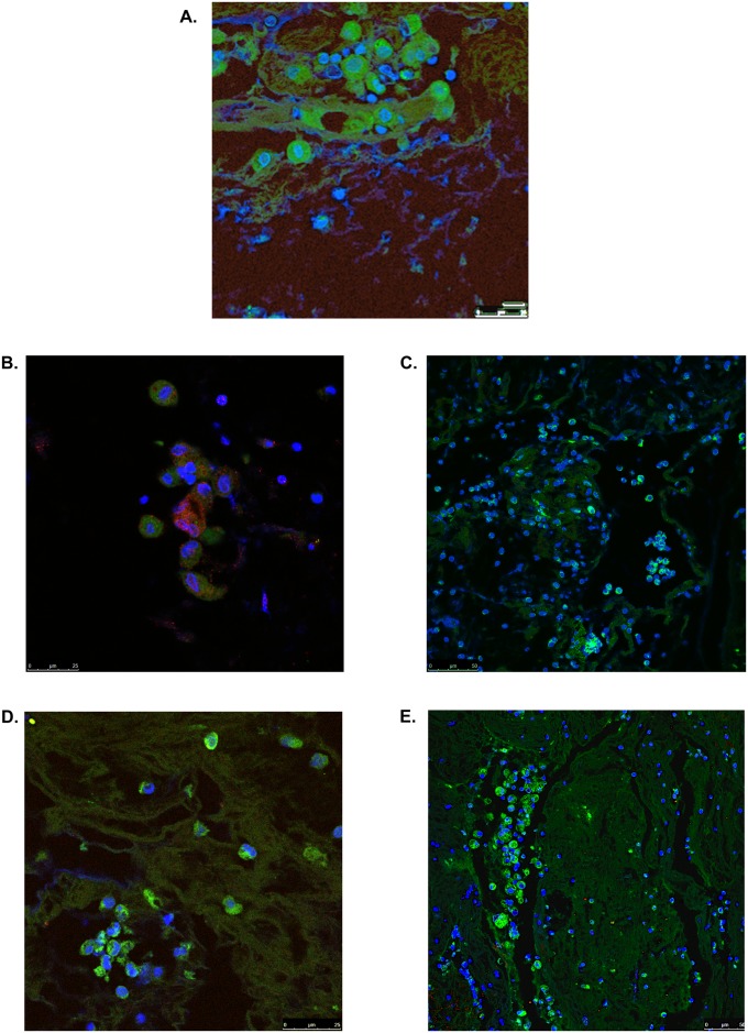
Figure 3.
Plastic bronchitis airway casts from children with Fontan physiology contain immune cells. Immunofluorescence (IF) confocal microscopy revealed positive staining for clusters of cells expressing (A) myeloperoxidase (green), indicative of neutrophils (original magnification, ×100; zoom factor 2.0); (B) eosinophilic peroxidase (red), which colocalized with CD4 (green) (original magnification, ×100; zoom factor 2.0); and (C) CD14 (green) and CD11b (red), indicative of macrophages (original magnification, ×40). (D) The presence of B lymphocytes was detected by CD20 (green) staining (original magnification, ×100; zoom factor 1.14). (E) CD4 staining (green) revealed clusters of eosinophils and lymphocytes and CD8 staining (red) was sparse. Samples were counterstained with 4′,6-diamidino-2-phenylindole (DAPI; blue) to visualize nuclei. Images are representative of at least one cast sample from four patients. The presence of T cells in cast samples was confirmed by immunohistochemistry (IHC) for CD3 (see Figure 2 and Figure E1). Representative IF and IHC of control slides that demonstrate antibody specificity and performance can be found in Figure 2 and in Figure E1.

