Figure 1.
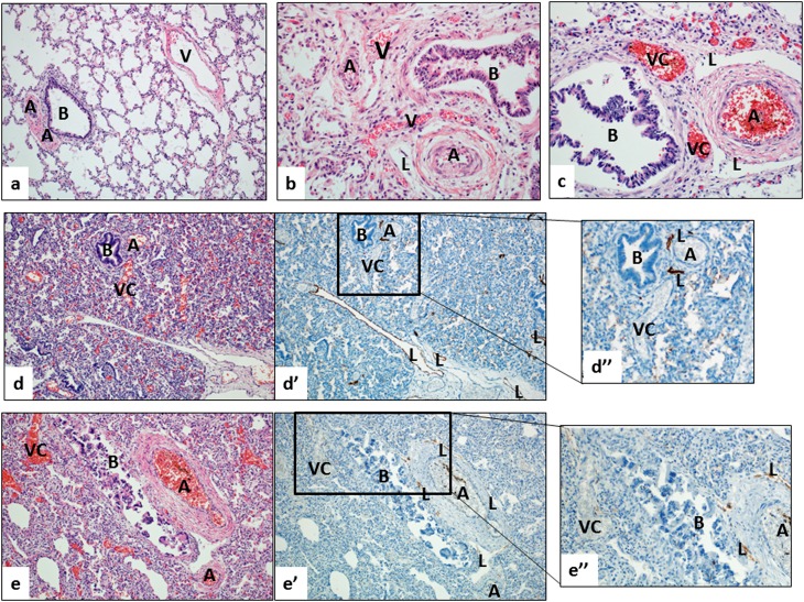
(a–c) Lung histology from young infants without respiratory disease (normal; a), with alveolar capillary dysplasia and misalignment of pulmonary veins (ACD/MPV; b), and bronchopulmonary dysplasia (BPD; c). (a) In the age-matched control lung, the pulmonary vein (V) is separated from the neighboring bronchoarterial bundle (B = bronchiole; A = pulmonary artery). Note the lack of prominent vessels other than the pulmonary arteries (A) near the airway (B). (b) In ACD/MPV, this normal vascular anatomy is disturbed, showing thin-walled and blood-filled vessels (“misaligned pulmonary veins,” V) that are abnormally located near pulmonary arteries (A) and bronchiole (B). (c) In severe BPD, prominent, thin-walled vascular channels with no elastic lamina (VC) are located near pulmonary arteries and airways, similar to the pattern seen in ACD (B = bronchiole; A = pulmonary artery; L = lymphatic vessel). Note that small and congested capillary network adjacent to VCs and surrounding airways and pulmonary arteries are seen in ACD and BPD lungs, but not in the lung of age-matched control. (d–e) The prominent blood-filled vascular channels (VC) in BPD lungs are not lymphatic channels. Serial step sections stained with hematoxylin and eosin (d, e) and D2-40 (d′, e′) show that the prominent vessels in the bronchoarterial bundles are not labeled with the lymphatic marker D2-40; this finding is further highlighted with a higher magnification (d′′, e′′). In contrast, typical lymphatic channels (L) show strong immunoreactivity with D2-40.

