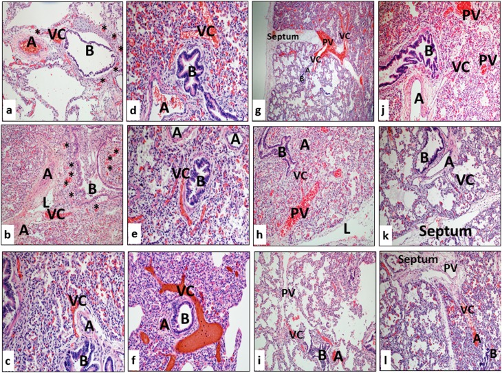
Figure 2.
Histologic patterns illustrating numerous and prominent blood-filled vascular channels (VC) within and adjacent to the bronchoarterial bundles in each of the lung samples from patients dying with severe bronchopulmonary dysplasia (A = pulmonary artery; B = bronchiole; L = lymphatic channel). Asterisks in panels a and b indicate congested dilated microvascular plexuses adjacent to pulmonary arteries (A), airways (B), and vascular channels (VC). See details in text.

