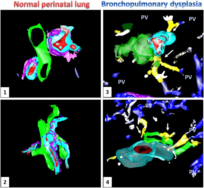
Figure 3.
Three-dimensional reconstruction of microscopic images from age-matched control (Panels 1 and 2) and bronchopulmonary dysplasia (BPD) (Panels 3 and 4) lungs. Prominent vascular channels (yellow) are present within the bronchoarterial bundles of BPD lungs (artery: red; connecting vessels: blue, pulmonary veins: blue; airway: green; lymphatic: pink; arterial muscular wall: aqua), but not in the control lungs. These vascular channels appear to course toward the vessels located within the interlobular septa (pulmonary veins, PV) and make contact with the microvessels surrounding the pulmonary arteries and airways (white arrows).

