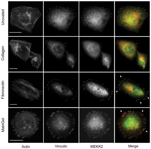
Fig. 1.
Cell attachment to matrix protein induces MEKK2 re-distribution. MDA-MB 231 cells seeded on glass coverslips that were either uncoated (first row), or coated with collagen (second row), fibronectin (third row), or Matrigel (fourth row) for 6 hours, fixed and stained for immunofluorescence analysis with anti-MEKK2 (green) or anti-vinculin (red) antibodies. Arrowheads indicate areas of MEKK2 localization to focal adhesions. Scale bar represents 20 μm. Images are representative of at least three independent experiments.

