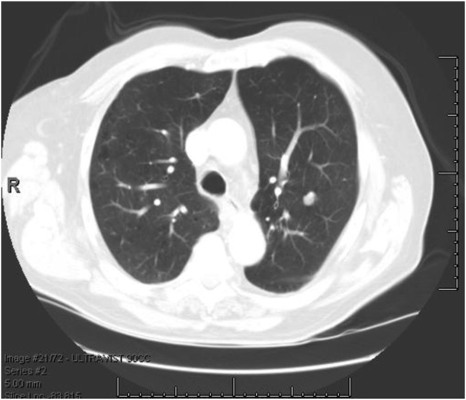
Figure 1.
Representative clinical vignette for a patient with a benign pulmonary nodule. Vignette 30: Mr. AD is an 82-year-old man and a former smoker. He was found to have a 10-mm, noncalcified nodule in the left upper lobe on chest CT. What is your best estimate for the probability that this nodule is malignant? CT = computed tomography.

