
Figure 5. AAQ1 competitively inhibits UBQLN2 binding to both full length and C-terminal TDP-43.
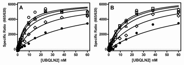
Various concentrations of AAQ1 and UBQLN2 were incubated with either full length TDP-43 (1.6 nM) (A) or C-terminal TDP-43 (261-414 aa, 4.6 nM) (B) for 2 hrs at room temperature followed by the addition of HTRF antibodies and subsequent ratiometric measurement of time resolved fluorescence as described in Section 2.5. Saturation binding isotherms for UBQLN2 binding to full length TDP-43 (A) or C-terminal TDP-43 (261-414 aa) (B) in the absence (○) or presence of 1 μM (□), 3.2 μM (△), 10 μM (◇), 32 μM (•) AAQ1. Global nonlinear regression fits of the data to a competitive inhibition model were obtained using Graph Pad Prism®. Data are the means of duplicate determinations from a single experiment that was repeated at least three times with similar results.