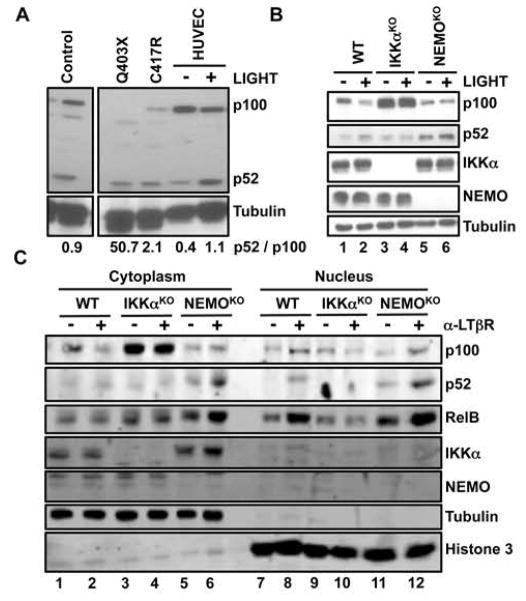
Fig. 1. The extent of p100 processing is enhanced in NEMO-ID cells and cells lacking NEMO.
(A) Lysates of PBMCs from patients with distinct NEMO mutations (Q403X and C417R), PBMCs from a healthy donor (Control) and HUVECs that were either untreated (−) or were stimulated with LIGHT for 8 hours (+) were analyzed by Western blotting with antibodies against the indicated proteins. Values reported here (in numbers beneath the blots) for the quantification of the ratio of the intensity of the p52 bands to the intensity of the p100 bands are arbitary units relative to the intensity of the tubulin bands. (B) WT, IKKαKO, and NEMOKO MEFs were either untreated (−) or were stimulated with LIGHT for 12 hours (+). Cell lysates were then analyzed by Western blotting with antibodies against the indicated proteins. (C) WT, IKKαKO, and NEMOKO MEFs were either left untreated (−) or were incubated with anti-LTβR antibody (α-LTβR) for 12 hours (+). Cytoplasmic and nuclear extracts were then prepared and analyzed by Western blotting with antibodies against the indicated proteins. Western blots are representative of three independent experiments.

