
Figure 1.
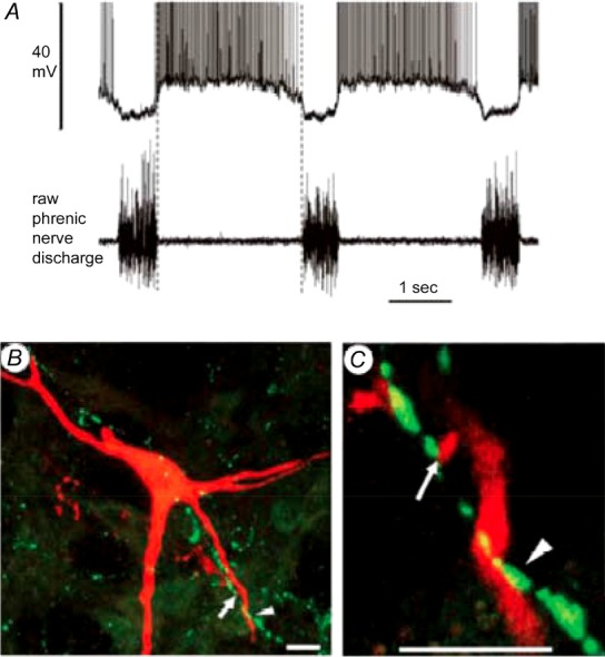
A, membrane potential and discharge trajectories of a decrementing expiratory (E-Dec) neuron in the VRC (upper trace) with corresponding phrenic nerve activity (lower trace). B and C, merged single slice confocal scans (1.8 μm thick) showing close appositions between DOR-immuno-reactive presynaptic terminal boutons and the dendrites (arrowhead) and labelled boutons (arrow) of the E-Dec neuron shown in A. Scale bars, 10 μm. Figure adapted with permission from Lonergan et al. 2003b, Fig. 2, panels O–Q.