Figure 3.

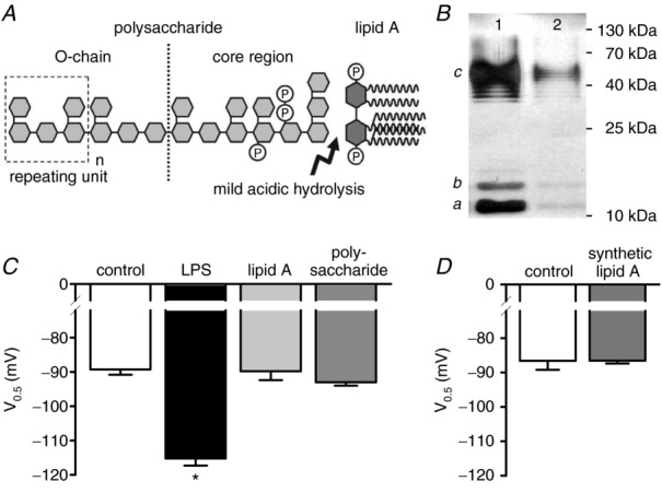
A, schematic chemical structure of lipopolysaccharide (LPS) (according to Rietschel et al. 1994). The arrow indicates the chemical bond, which is cleaved by mild acetic acid hydrolysis, resulting in a separation of the polysaccharide moiety containing the O-chain from the hydrophobic lipid A. B, 7.5 μg of untreated (lane 1) or hydrolysed (lane 2) LPS were subjected to 12% SDS-PAGE and silver-stained. The typical staining pattern of S-type LPS is characterized by intermediates of the full-length LPS representing lipid A with the core oligosaccharide only (a), lipid A and core with one repeating unit (b), and lipid A and core with different numbers of the repeating unit (c) (according to Palva & Makela, 1980). Because only intact LPS can be separated and visualized by this method, the reduction in staining intensity indicates 90% hydrolysis of LPS. C, mean V0.5 recorded under control conditions (open bar, n = 11), in the presence of 50 μg ml−1 LPS (black bar, n = 11) and in the presence of degradation products derived from 50 μg ml−1 LPS exposed to mild acidic hydrolysis (lipid A: light grey bar, n = 10; polysaccharide: dark grey bar, n = 10). Only the intact LPS molecule affects V0.5 of IhHCN2. D, compared to control (open bar, n = 6), V0.5 of IhHCN2 is not modulated by 10 μg ml−1 synthetic lipid A (grey bar, n = 10). *P < 0.05 versus control.
