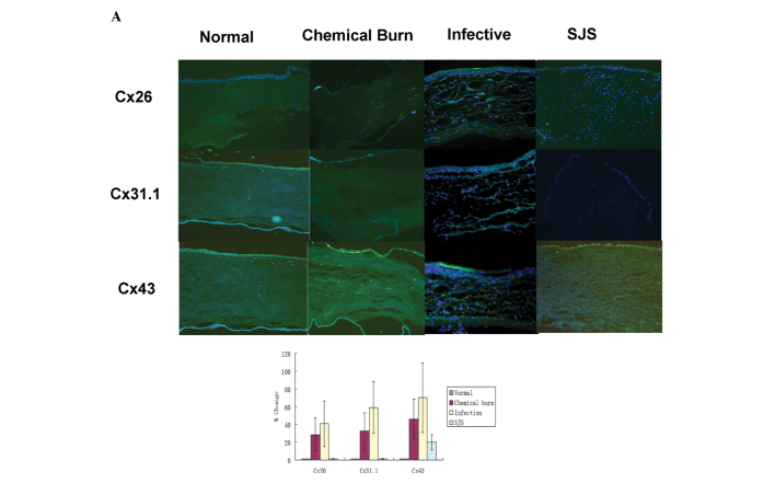
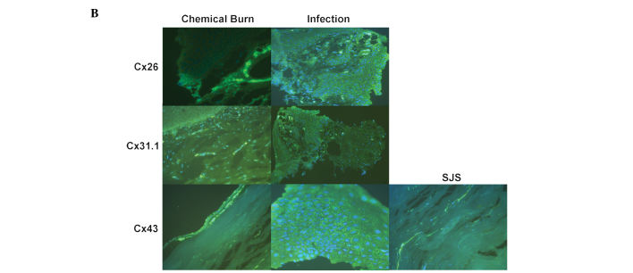
Figure 3.
Immunohistochemical expression of Cx26, Cx31.1 and Cx43 in human corneas. Nuclei of corneal cells are stained blue (DAPI). Positive immunolabelling appears green (Cy2). (A) Expression of Cx26, Cx31.1 and Cx43 was quantified and the results are expressed in a bar graph. *P<0.05 vs. normal cornea. Immunostaining for Cx26 and Cx31.1 was upregulated in chemically burned corneas, infected corneas and allograft failure corneas. Immunostaining for Cx43 was upregulated in all the diseased cornea groups. (A), magnification, ×100; (B), magnification, ×400. Cx, connexin; SJS, Stevens-Johnson syndrome.


