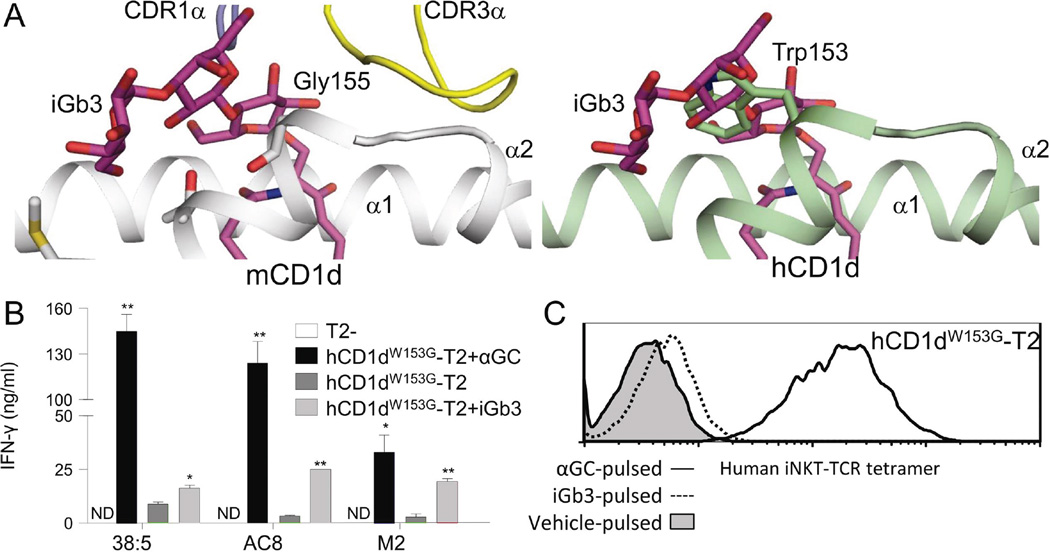
Figure 4.
Tryptophan W153 in hCD1d is responsible for human-mouse species differences in iGb3 presentation. (A) The crystal structure of iGb3-mCD1d complexed with iNKT TCR (left, from Pellicci et al., 2011) and a superposition of the iGb3 headgroup on hCD1d (right) are shown, suggesting that W153 in hCD1d could sterically hinder the iNKT TCR-induced flattening of the iGb3 headgroup. (B) IFN-γ release by three human iNKT clones (38:5, AC8 and M2) in response to hCD1dW153G-T2 cells that were pulsed with either black αGC (black columns), vehicle (dark grey columns) or iGb3 (light grey columns). Data were generated in parallel with Figures 1C and 3B and are shown as mean ± SEM of XXX samples/repliates, representative of/pooled from XXX experiments. *p<0.05, **p<0.01, compared with vehicle-pulsed hCD1dW153G-T2, paired t-test. ND: Not detected. (C) Staining of αGC-pulsed (black line histogram), iGb3-pulsed (dotted line histogram) and vehicle-pulsed (grey-filled histogram) hCD1dW153G-T2 cells with fluorescent-conjugated human iNKT TCR tetramer. Data were generated in parallel with Figure 3C, and are representative of three independent experiments.

