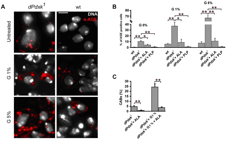
Figure 7. dPdxk1 mutant brains exhibit higher frequencies of AGE-positive cells than wild type (wt) brains.
(A) Examples of cells stained with a rabbit anti-human AGE antibody. Scale bar, 5 µm. (B) Frequencies of AGE-positive cells in wild type (wt) and dPdxk mutant brains exposed to different glucose (G) concentrations with or without ALA or PLP; bars represent the mean frequencies of AGE-positive cells (±SE) obtained by examining at least 300 cells/brain in 4 brains. At all glucose concentrations untreated dPdxk1 mutant brains have a significantly higher frequency of AGE-positive cells than wild type controls and either ALA or PLP treated dPdxk1 brains; (C) ALA reduces the frequency of CABs induced by dPdxk1 or dPdxk1 plus 1% G. Each bar in the graph represents the mean frequency of CABs (±SE) obtained by examining at least 600 metaphases form at least 6 brains. * and **, significantly different in the Student t test with p<0.03 and p<0.001, respectively.

