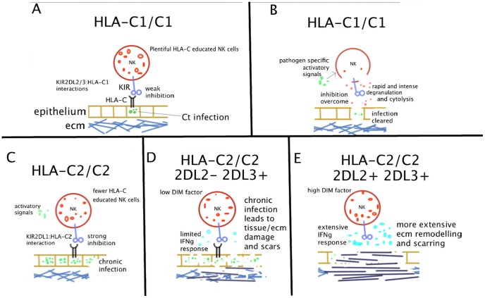
Figure 4. Model of NK cell mediated scarring in trachoma.
(A) An HLA-C1/C1 NK cell interacts with HLA class I molecules on a Ct infected epithelial cell via interactions between KIR2DL2/L3 and HLA-C1. HLA-C educated NK cells represent a greater proportion of the total NK repertoire in HLA-C1 homozygotes, compared to those who carry HLA-C2 (B) The inhibitory signals are overcome by loss of the inhibitory HLA-C molecule and/or by activatory signals, which may be from intracellular or extracellular pathogen derived moieties. The NK cytotoxic response is triggered and the target cell is lysed, which culminates in effective resolution of the infection. (C) An HLA-C2/C2 NK cell interacts with MHC class I molecules on Ct infected epithelial cell via interactions between KIR2DL1 and HLA-C2. A smaller proportion of the NK cell repertoire is HLA-C educated in this setting. Cytotoxic and IFNγ responses are less likely to be triggered and responses are less intense than in HLA-C1/C1 individuals. Chronic infection is established (D) KIR2DL2 −, KIR2DL3 + individuals have a low DIM factor and NK cell release of IFNγ is more limited. Chronic infection leads to some damage to epithelium and extracellular matrix (ECM) but anti-fibrotic properties of IFNγ are maintained. (E) KIR2DL2/KIR2DL3 heterozygous individuals have a high DIM factor and NK cells release larger quantities of IFNγ. Chronic infection is coupled with pathologically high levels of IFNγ. HLA-C1/C2 genotypes confer intermediate risks that do not significantly differ from the homozygous individuals. Relative risk of scarring was increased in KIR2DL2 homozygotes, but this was not significant.

