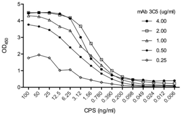
Figure 2. Detection of purified CPS by antigen-capture ELISA.

mAb 3C5 was used in the capture phase of the ELISA at the concentrations listed. Following a wash and blocking step, purified CPS was serially diluted across the microtiter plate at the concentrations listed. The wells were then washed and HRP-labeled mAb 3C5 was used in the indicator phase to detect captured CPS. The ELISA was performed in triplicate and mean values are plotted.
