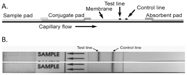
Figure 3. Prototype Active Melioidosis Detect (AMD) LFI.

(A) Schematic of LFI components. (B) B. pseudomallei strain Bp82 colony grown on an agar plate was picked and suspended in 2 drops of lysis buffer. The lysate was added to the sample pad followed by three drops of LFI chase buffer (top LFI). The LFI was imaged following a 15 min run time. The same test condition were used with a colony of E. coli (bottom LFI).
