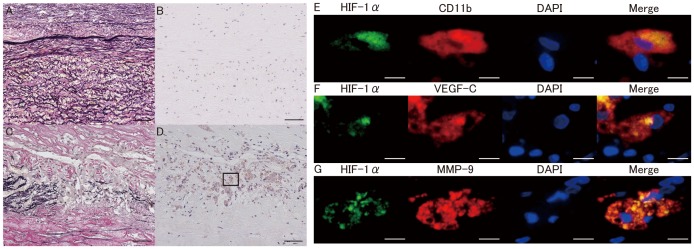
Figure 3. HIF-1 expression in normal aorta and AAA.
Elastica van Gieson staining of the intima/media in normal aorta (A) and abdominal aortic aneurysm (AAA) wall (C). Immunohistochemistry for HIF-1α in normal aorta (B) and AAA (D). Nuclear and cytoplasmic expression of HIF-1α was observed in intima/media in AAA (D). E, Double immunofluorescence staining for HIF-1α (green), CD11b (red), DAPI (blue), and the merged image of the outlined in D. F, Double immunofluorescence staining for HIF-1α (green), VEGF-C(red), DAPI(blue), and the merged image of the outlined in D. G, Double immunofluorescence staining for HIF-1α(green), MMP-9 (red), DAPI (blue), and the merged image of the outlined in D. Expression of HIF-1α was increased in CD11b positive macrophages. VEGF-C and MMP-9 were expressed in the HIF-1α–positive macrophages. Scale bars indicated 50 µm (A–D) and 10 µm (E–G).

