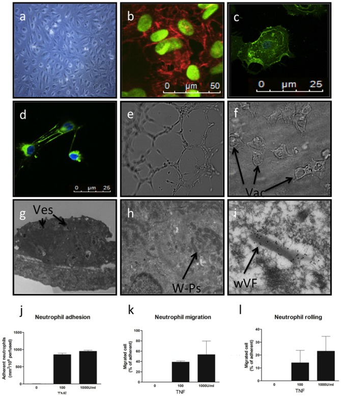
Figure 1. The BOEC is functionally a mature endothelial cell.
a) Phase microscopy of BOECs in culture demonstrates typical endothelial “cobblestone” morphology. Confocal microscopy of BOECs with nuclear counterstaining. b) CD31 (red), c) CD146 (green), d) fibronectin (green). e) Network formation in matrix gel, f) vacoulised BOECs in fibronectin/collagen matrix. g) Electron microscopy (EM) of whole BOEC showing extensive vesicles (Ves), h) EM close up of W-P bodies, i) EM of W-P bodies with immunogold staining for vWF. j-l) Neutrophil rolling adhesion and transmigration after stimulation with TNF in a flow-based assay.

