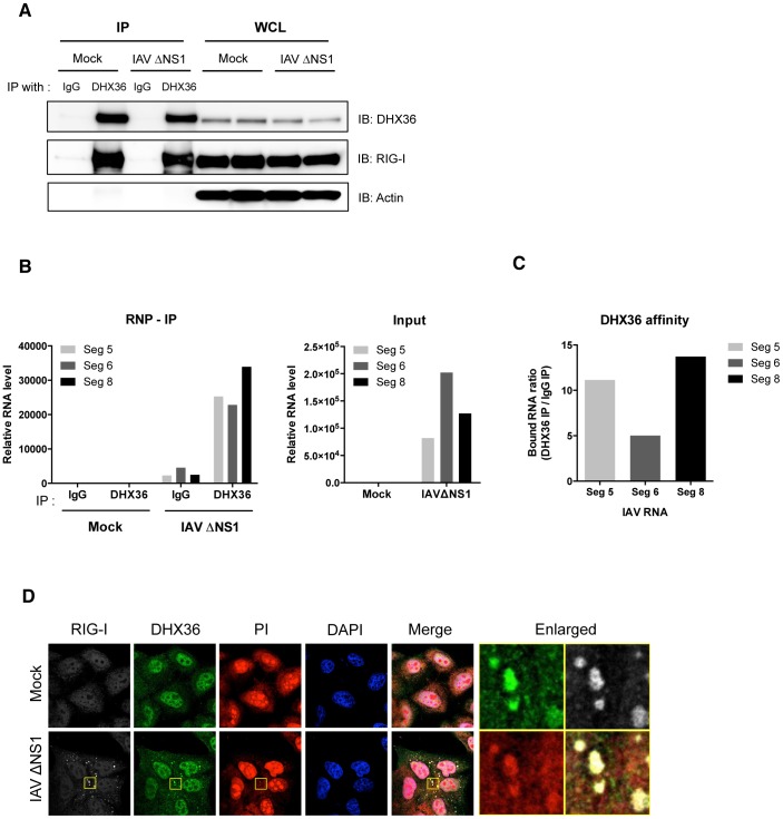
Figure 3. DHX36 recognizes viral RNAs.
(A–C) HeLa cells were mock-treated or infected with IAVΔNS1. After 12 h infection, cells were collected and lysed for RNA-IP analysis. After pull-down with indicated antibodies, RNA was recovered from the RNP complex. The IP efficiency was confirmed by Western blot analysis by indicated antibodies (A). The level of RNAs bound to RNP complex as well as input RNAs was measured by real-time qPCR with indicated probes (B). DHX36 affinity was evaluated by calculating the ratio of RNAs from DHX36 IP and IgG IP (C). (D) HeLa cells were mock-treated or infected with IAVΔNS1. After 12 h infection, cells were fixed and stained for the indicated proteins. Cytoplasmic dsRNAs were detected by PI staining. Nuclei were visualized by DAPI staining. The images were taken by confocal microscopy. High magnification images of the square area are shown (Enlarged).

