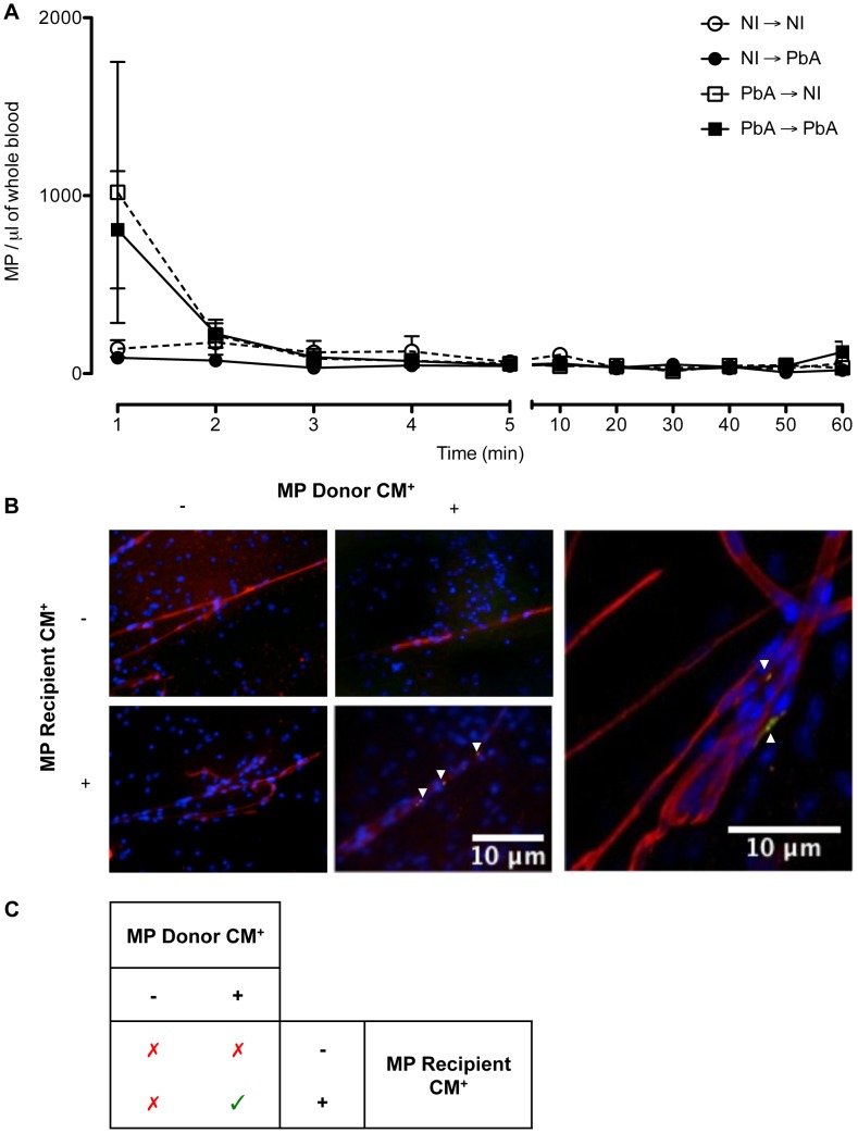
Figure 5. Detection, clearance, and tissue distribution of MP following adoptive transfer.
(A) Clearance of transferred MP within circulation following adoptive transfer. The presence of transferred PKH67-labeled MP was detected via flow cytometry in the blood of recipient mice. MP were detectable immediately following intravenous injection; healthy recipients (circles) cleared the MP quicker than PbA infected recipients (squares). Data are mean ± SEM from n = 3 per group. (B) Microparticles localised in brain microvessels of CM+ recipient mice. Smears were prepared from healthy and CM+ mice, recipients of PKH67-labelled MP (green) purified from healthy or CM+ donor mice (n = 3). MP were activated in vitro with calcium ionophore (A23187), a known vesiculating agonist, labelled, intravenously transferred and allowed to circulate for 1 h. Brain smears were fixed and counter-stained with Texas Red Lectin and DAPI to identify vessels (red) and nuclei (blue), respectively. MP from CM+ donors can only be detected in the microvessels of CM+ recipient mice (arrows). Imaged on Olympus IX71. Insert: MP from CM+ donor lodged in CM+ brain vessel, imaged using oil immersion ×60 on Olympus FluoView FV 1000 confocal microscope. Arrows indicate MP lining the endothelium amongst other cells and trapped in birfurcation of vessels. (C) Summary of MP localisation in cerebral vessels. MP from CM+ donors can only be detected in the microvessels of CM+ recipient mice, as indicated by green tick.

