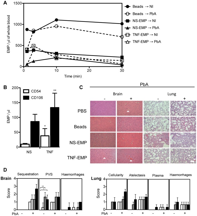
Figure 6. Transferred EMP generated in vitro induce CM-like pathology in healthy mice.
(A) Detection of PKH-67 labelled EMP in the blood of recipient mice following transfer. Purified EMP (400×103/mouse) from TNF stimulated and resting mouse brain microvascular endothelial cell line B3 harvested in vitro, were transferred into PbA infected and healthy recipient mice. Clearance of fluorescently labeled EMP from the blood was monitored by flow cytometry. Latex beads (400×103/mouse, control) can be detected in constant circulation up to 30 minutes post transfer. Data represent the mean from n = 3. Closed circle/solid line indicates beads transferred into healthy mice, open circle/interrupted line indicates beads transferred into infected mice, closed square/solid line indicates NS- EMP transferred into healthy mice, closed triangle/solid line indicates NS-EMP transferred into infected mice, open square/interrupted line indicates TNF-EMP transferred into healthy mice and open triangle/interrupted line indicates TNF-EMP transferred into infected mice. (B) Detection of CD54 and CD106 on in vitro generated MP derived from mouse brain microvascular endothelial cells. Graph shows the expression of CD54 (white bar) and CD106 (black bar) on resting and TNF-stimulated EMP. Data are mean ± SD, *p<0.05. The numbers of CD54+ and CD106+ EMP in the supernatant increased following TNF-stimulation (mean ± SEM MP/µL; CD54: 9.850±1.064 to 38.050±9.925; 74.11% CD54+ shift CD106: 86.183±9.981 to 133.250±19.860; 35.32% CD106+ shift). (C) Representative haematoxylin-eosin brain and lung sections from healthy and PbA infected mice treated with PBS or EMP (n = 3) on day 6 p.i. Morphometric analysis reveals that EMPs induce significant pathology in healthy brain and lung. In the brain, the arrows heads indicate areas of engorged vessels and haemorrhage. In the lung, multifocal lesions, abnormal cellularity in the alveolar septa, plasma in alveoli and haemorrhage. Treatment with EMP in healthy mice induces haemorrhage in the brain (white arrow head) and increased cellularity (white arrow head) and atelectasis (white arrow) in the lung of recipient mice. Such pathology is not observed in the mice treated with PBS or beads alone. Scale bar (bottom left) indicates 40 µm. (D) Effects of in vitro generated EMP on healthy and PbA infected brain and lung in the following treated groups: PBS (white bar), beads (light grey), NS-EMP (dark grey) and TNF-EMP (black). Representative Haematoxylin-Eosin images (n = 3) per group were scored for histopathological signs (Table 3).

