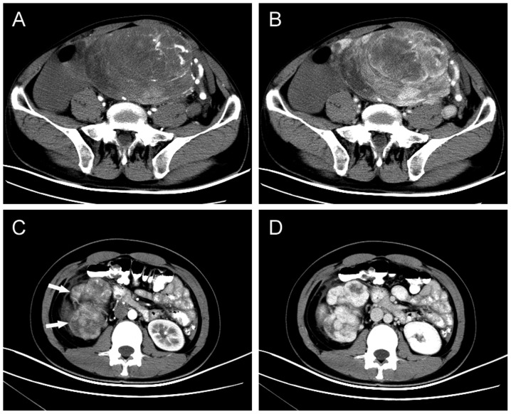
Figure 1.
Typical imaging manifestations of abdominopelvic SFTs. (A) Arterial-phase and (B) venous-phase axial contrast-enhanced CT scan showing a well-defined, intensely enhancing mass in the pelvis with central nonenhancing areas. (C) Lobulated SFTs (indicated by arrows) of the retroperitoneum in a 21-year-old female. (D) Axial contrast-enhanced CT scan, obtained in the arterial phase, revealing a well-defined hypervascular mass with intense enhancement. SFT, solitary fibrous tumor; CT, computed tomography.

