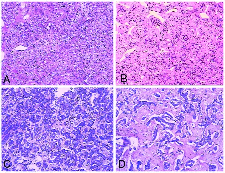
Figure 4.
Hematoxylin and eosin stained sections. (A) SFT consists of tightly packed round to fusiform cells with indistinct cytoplasmic borders which are arranged around a vessel. Dimensions, 10×10 cm. (B) The vessels formed a continuous, ramifying vascular network. The vessels divide and communicate with small or minute vessels which have been partly compressed and obscured by the surrounding cellular proliferation. Typically, the dividing sinusoidal vessels have an ‘antler-like’ configuration. Dimensions, 10×10 cm. (C) Malignant SFT with heightened cellularity. The tumor also demonstrates marked pleomorphism with a high level of mitotic activity (>4 mitotic fields/10 high power fields). Dimensions, 10×10 cm. (D) SFT tumor cells arranged randomly in a ‘patternless pattern’. The SFT presented marked hyalinization and had characteristic artifactual ‘cracks’ between the cells and collagen. Dimensions, 10×20 cm. SFT, solitary fibrous tumor.

