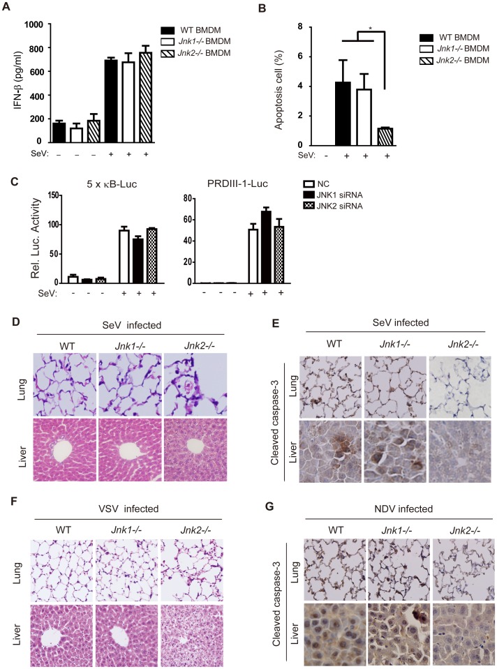
Figure 7. JNK2 protects mice against virus-induced organ injury.
(A) BMDMs from wild type, Jnk1−/− or Jnk2−/− mice were treated with or without SeV (MOI = 1) for 18 hours and then IFN-β production was determined by ELISA. Data are presented as means±SD (n = 3). (B) BMDMs from wild type, Jnk1 −/− or Jnk2 −/− mice were treated with SeV (MOI = 1) for 48 hours, followed by Annexin-V staining analysis. Cells undergoing apoptosis (Annexin-V positive and PI negative) were quantified by flow cytometry. Data are presented as means±SD (n = 3). (C) The indicated siRNAs were transfected into HEK293 cells together with NF-κB-Luc reporter or PRDIII-1-Luc reporter plasmids and 24 hours later, cells were infected with or without SeV (MOI = 1). A luciferase assay was performed 12 hours post-infection. Data are presented as means±SD (n = 3). (D and E) Wild type, Jnk1−/− or Jnk2−/− mice were intranasally challenged with SeV (107 PFU/g mouse weight). Two days later, lungs and livers were harvested for histochemistry analysis by H&E staining and immunohistochemistry analysis by detecting cleaved caspase-3 staining. (F and G) Mice were infected with VSV or NDV as described in Figure 6. Two days later, lungs and livers were harvested for histochemistry analysis by H&E staining and immunohistochemistry analysis by cleaved caspase-3 staining. Tissue sections were visualized by microscopy (20× objective for lung, 40× objective for liver).

