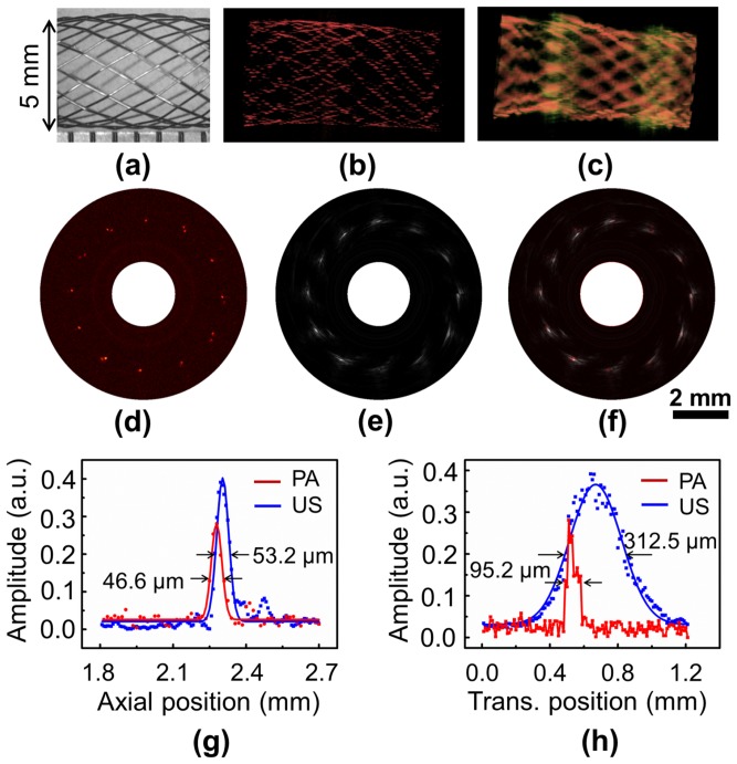
Figure 4. OR-PAT of an iliac/common femoral artery stent.
(a) Optical microscopic image of the imaged sent segment. (b) Representative 3D photoacoustic and (c) ultrasonic images. (d) Photoacoustic, (e) ultrasonic, and (f) fused B-scan images of a cross section of the stent. (g) Axial and (h) transverse photoacoustic (red) and ultrasonic (blue) signal profiles of a wire junction at 2 o′clock in (f). PA, photoacoustics; US, ultrasonics; Trans., transverse.

