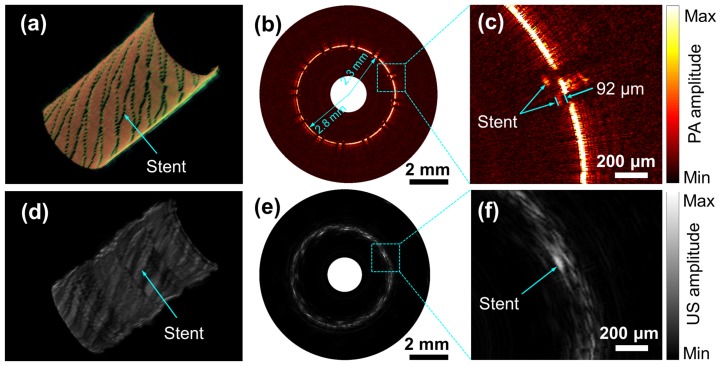
Figure 5. Photoacoustic and ultrasonic imaging with OR-PAT of a stent deployed in a plastic tube.
Three-dimensional cut-away (a) photoacoustic and (d) ultrasonic images. Representative (b) photoacoustic and (e) ultrasonic B-scans. Enlarged photoacoustic (c) and ultrasonic images corresponding to the dash boxes in (b) and (e).

