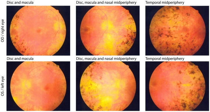
Figure 3. Fundus photographs from patient 121–385 (male, 43 years old).
The patient shows the representative phenotype of this mutation with waxy pallor of the optic disc, retinal arteriolar attenuation, granularity of the macula and intra retinal pigment around the midperiphery in a bone spicule or clumped configuration.

