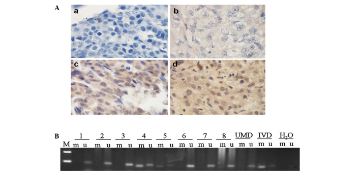
Figure 1.
Immunohistochemical staining and methylation of BRCA1 in sporadic EOC tissues. (A) Immunohistochemical staining of sporadic EOC specimens (magnification, ×1000): (a) Negative stain; (b) weak positive stain; (c) moderately positive stain; (d) strongly positive stain. (B) Methylation of BRCA1. MSP was performed on bisulfite-treated DNA from ovarian cancer cells. MSP results from nine representative patients are shown. The DNA bands in the u-labeled lanes indicate PCR products that were amplified with primers recognizing the unmethylated promoter sequence. The DNA bands in m-labeled lanes represent the products that were amplified with methylation-specific primers. The DNA from the normal lymphocytes served as the control for UMD and IVD served as the control for methylated DNA. H2O was used as a template for the negative control. M, marker; UMD, unmethylated DNA; IVD, in vitro methylated DNA; BRCA1, breast cancer susceptibility gene 1; EOC, epithelial ovarian carcinoma; MSP, methylation-specific polymerase chain reaction.

