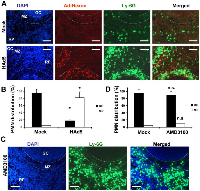
Figure 3. Specific Ly-6G+ leukocyte retention in the splenic marginal zone occurs in response to Ad, but not AMD3100.
(A) Distribution of Ly-6G+ cells (green, FITC-labeled anti-Ly-6G antibody) on sections of spleen harvested from mice that were injected with either Ad or saline (Mock panels) 2 hours after treatment. The splenic marginal zone (MZ), located between the germinal centers (GC) and red pulp (RP) anatomical compartments, is outlined with dotted lines and was defined based on section staining with DAPI (blue). MZ macrophages trap Ad particles from the blood (Ad-Hexon antibody staining, red). Representative panels are shown. N = 5. Sections at three depth levels were obtained from each mouse. (B) Quantitative representation of PMN distribution on sections of the spleen 2 hours after virus challenge. Ly-6G+ cell (PMN) distribution was analyzed on at least 4 consecutive sections of the spleen, cut at three depth levels from each individual mouse. The pictures of spleen sections with average PMN cell densities were taken and up to 200 total PMN cells were counted and assigned to MZ or RP compartments based on their actual localization. For each experimental condition, the data were combined and averaged from three to 5 individual mice. * - P<0.05. Mock – mice injected with saline. (C) Distribution of Ly-6G+ cells (green, FITC-labeled anti-Ly-6G antibody) on sections of spleen harvested from mice that were injected with small molecular drug AMD3100 2 hours after treatment. The splenic marginal zone (MZ), located between the germinal centers (GC) and red pulp (RP) anatomical compartments, is outlined with dotted lines and was defined based on section staining with DAPI (blue). Representative panels are shown. N = 5. Sections at three depth levels were obtained from each mouse. (D) Quantitative representation of PMN distribution on sections of the spleen 2 hours after the mouse was challenged with small drug AMD3100. The cellular localization was assessed and analyzed as described in (B). n.s. – not statistically significant, compared to corresponding control group. RP – red pulp; MZ – marginal zone.

