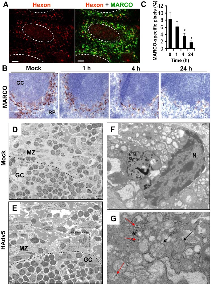
Figure 7. MARCO+ marginal zone cells are eliminated from the spleen after sequestering Ad from the blood.
(A) Distribution of Ad particles (stained with anti-Hexon Ab, red) and MACRO+ cells (stained with anti-MARCO Ab, green) on spleen sections 1 hour after intravascular virus injection. Co-localization of virus particles with MARCO+ staining appears as yellow. The anatomic borders of splenic germinal centers are depicted with dotted lines. Scale bar is 50 µm. Representative images are shown. N = 25. (B) Immunohistochemical analysis of MARCO+ marginal zone macrophages on sections of spleen at different times after Ad administration. Spleens of mice injected with the virus were harvested at indicated times and stained with MARCO-specific antibodies. Sections were counter-stained with hematoxylin to visualize splenic anatomical compartments. Mock – spleen sections were prepared from mice injected with saline only. GS – germinal center. RP – red pulp. Representative fields are shown. N = 5. (C) Quantitative representation of MARCO+-specific staining on splenic sections of mice after Ad administration. N = 4 per experimental group per time point. Marker-specific pixels (brown staining) were quantified using MetaMorph software on low-power images of spleen sections with average distribution of MARCO-specific staining collected from three sections and at three depth levels. * - P<0.05. Error bars represent standard deviation of mean. (D–G) Transmission electron microscopy analysis of ultra-thin sections of spleens harvested from saline-injected mice (D) or Ad-injected mice (E) 4 hours after the virus injection. MZ – marginal zone; GC – germinal center. The anatomic borders of germinal centers are depicted with a punctuated line. (F and G) Punctuated rectangle depicts a marginal zone macrophage that contains Ad particles (red arrows on G) and morphologically distorted mitochondria (F and black arrows on G). N – nucleus. Representative images of virus-containing cells in the marginal zone are shown.

