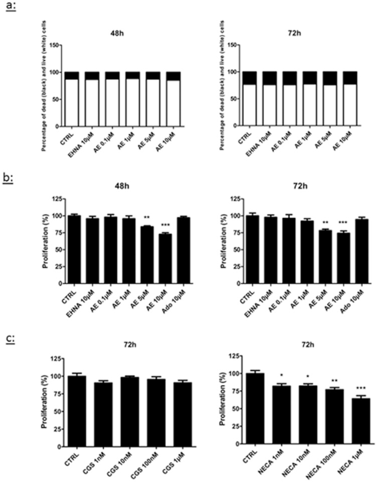
Figure 1. Effect of adenosine on LEC viability and proliferation.
Cell viability (a) and proliferation (b–c) were evaluated in HMVEC-dLy ( = LEC) cultured for 48 h (left panel) or 72 h (right panel) in medium containing 2% FBS (CTRL). Cells were treated with EHNA alone (EHNA 10 μM), EHNA with different concentrations of adenosine (AE), the A2a agonist CGS21680 or the A2b agonist NECA. For cell viability (a), results are expressed as percentage of dead cells (black) and living cells (white). Cell proliferation (b–c) was measured with a CyQANT assay. * p<0.05 vs control (CTRL), *** p<0.001 vs CTRL. Data are expressed as mean ± SEM (n = 3).

