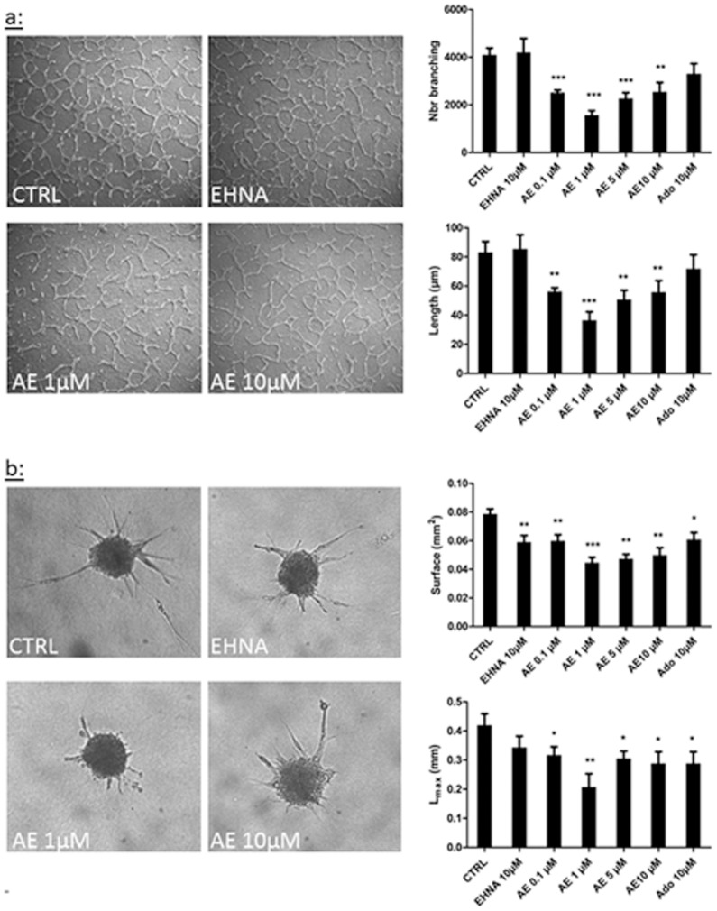
Figure 4. Effect of adenosine on LEC tube formation.
Tube formation was assessed in two in vitro models, the tubulogenesis assay (a) and the spheroid assay (b). HMVEC-dLy were cultured in 2% FBS medium (CTRL) and treated or not with EHNA alone (EHNA 10 μM) or EHNA with different concentrations of adenosine (AE). Quantification of tube formation (a) and cell migration (b) was performed by a computerized method on pictures taken after 24 h of culture. The parameters measured are: the tubes branching (branching), the length of tube (length), the surface occupied by tube (surface), and the maximal length of tube (Lmax). * p<0.05 vs CTRL, **p<0.01 vs CTRL, *** p<0.001 vs CTRL. Each experiment was performed three times and representative pictures are shown. Data are expressed as mean ± SEM.

