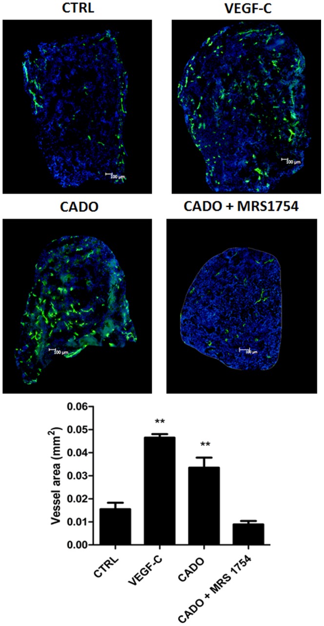
Figure 5. Effects of adenosine on lymphangiogenesis in the in vivo model of collagen sponge.

Sponges were soaked with PBS as control (CTRL), with VEGF-C (1 ng/ml) as positive control (VEGF-C), with 20 μl CADO (3 ng/ml), a stable analog of adenosine, or with CADO in presence of the A2b antagonist MRS1754 (2.4 mg/ml). Sponges were implanted between the two skin's layers of ear's mice for 3 weeks. Every other day, PBS or MRS1754 were injected in the apex of the ear. Sponge sections were stained with an anti-Lyve-1 antibody to detect lymphatic vessels (green) and Dapi to detect cell nucleus (blue). The graph corresponds to computerized quantification of the surface occupied by lymphatics (vessel area). Data are expressed as mean ± SEM (n = 6). ** p<0.01 vs CTRL.
