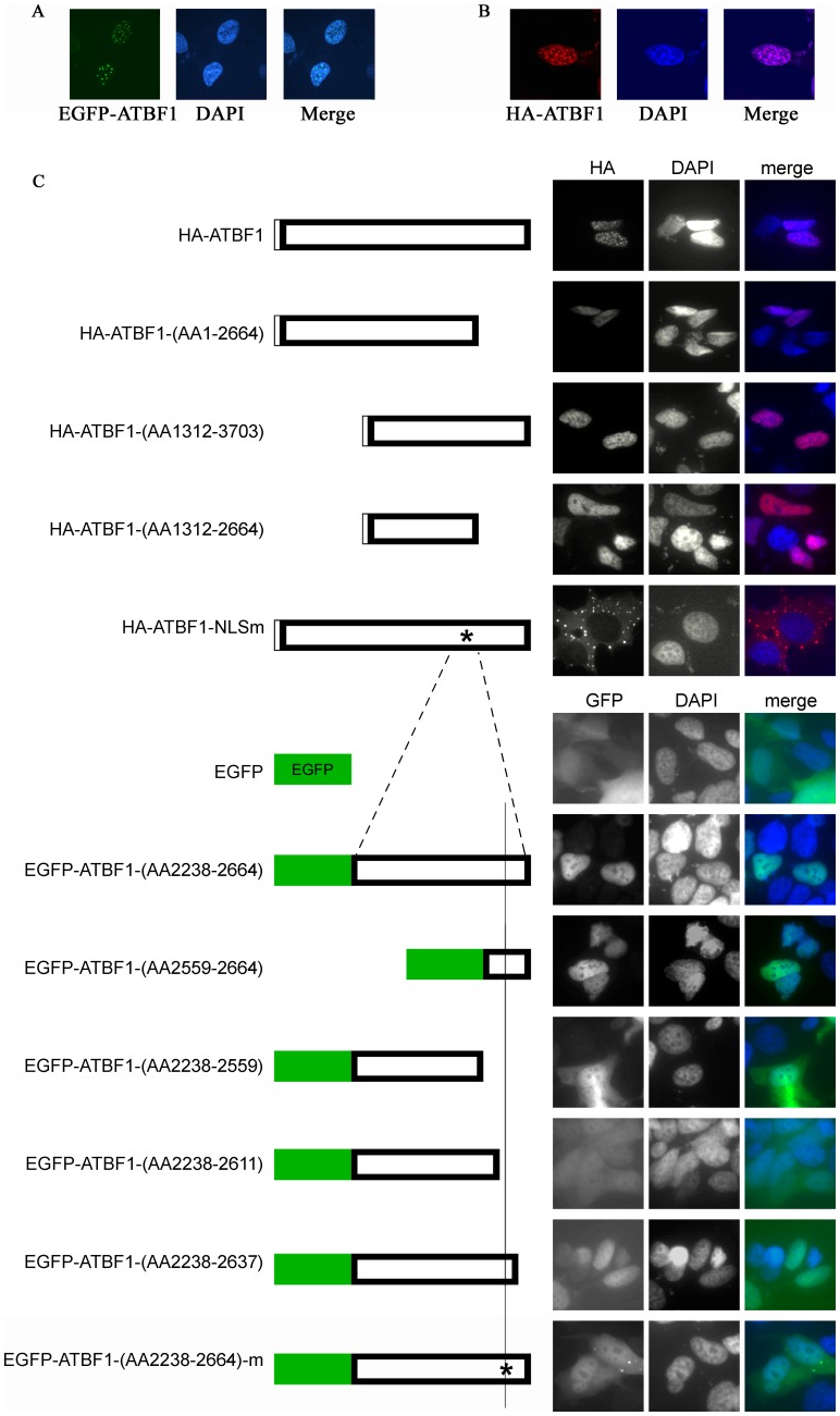
Figure 1. Detection of nuclear body (NB)-like ATBF1 dots in the nucleus and definition of the ATBF1 nuclear localization signal (NLS).
A, B. ATBF1 concentrates to form NB-like dots in the nucleus, as detected by immunofluorescent microscopy in 22Rv1 cells expressing EGFP-fused ATBF1 (A) or HA-tagged ATBF1 (B). C. Identification of the NLS for ATBF1 by deletion mapping, mutation and immunofluorescent microscopy. Each box at left represents a fragment of ATBF1 or the full length ATBF1 (wildtype or mutant as indicated by *) attached with the EGFP or the HA tag. Each fragment box is aligned to full length ATBF1 to indicate its relative location in ATBF1. Images at right indicate the subcellular localization of a fragment or full length ATBF1, with the nucleus shown by DAPI staining. Residue numbers are based on the ATBF1-A protein sequence (NCBI access number NP_008816).

