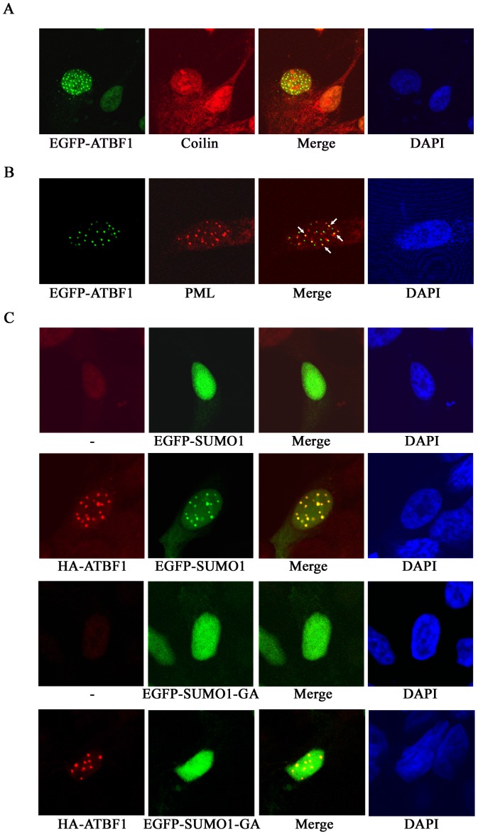
Figure 2. Association of ATBF1 with PML nuclear bodies (PML NBs) and SUMO1 in the nucleus, as detected by immunofluorescent microscopy in 22Rv1 cells.
A, B. ATBF1 dots are not associated with Cajal bodies (A) but partially overlap with a subset of PML NBs (arrows) (B). ATBF1 dots were visualized by EGFP (green), and Cajal bodies and PML NBs by an anti-Coilin or anti-PML antibody, respectively (red). C. Co-localization of ATBF1 and SUMO1. ATBF1 dots were detected by an anti-HA antibody (red), and wild type and mutant SUMO1 proteins by EGFP (green). While wildtype SUMO1 is co-localized with ATBF1 dots, the mutant form (EGFP-SUMO1-GA) is not. Nuclei are shown by DAPI counterstaining.

