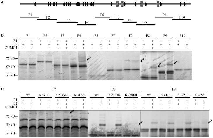
Figure 4. Identification of SUMOylation sites of ATBF1.
A. Location of 10 ATBF1 fragments (F1-F10) relative to the full-length ATBF1 protein (top) for the in vitro SUMOylation assay. In the schematic for full-length ATBF1, potential functional domains, including zinc fingers (ovals) and homeodomains (rectangles), are shown based on previous predictions by Miura et al. [1]. B. Detection of SUMOylated ATBF1 fragments in vitro. For each fragment, SUMO1 was present (+) or absent (-) in the reactions. Arrows point to SUMOylated ATBF1 fragments. C. Identification of lysine residues that are SUMOylated in the 3 ATBF1 fragments. In vitro SUMOylation assay was performed for ATBF1 fragments with different lysine mutants. Arrows indicate the disappearance of SUMOylated ATBF1 peptides in the K2349R, K2806R and K3258R mutations of the 3 fragments.

