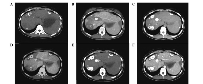
Figure 1.
Three-year follow up CT images of one patient (no. 4) who did not receive any specific therapy. (A and B) First plain CT image showed multiple hypoattenuating coalescent nodules in the peripheral region of the liver with capsular retraction and mild calcification. (C and D) Second CT (one-year later) showed no change in tumor size, but an increase in calcification. (E and F) Third CT examination (two-years later) reconfirmed the regression of the tumor with the augmentation of calcification replacing the previous tumor cells. CT, computed tomogrphy.

