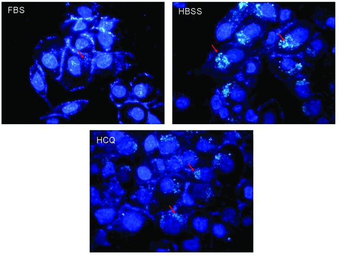
Figure 4.
Specific GFP-LC3 combined fluorescence microscopy in cervical cancer SiHa cells treated with HBSS or HCQ. Cells were treated with 20 μmol/l HCQ and starvation for 12 h and stained with specific green fluorescent protein-LC3. Cell morphology was observed by fluorescence microscopy (red arrows indicate autophagosomes or LC3 accumulation). FBS, fetal bovine serum; HCQ, hydroxychloroquine; HBSS, Hank’s balanced salt solution.

