
Fig. 1.
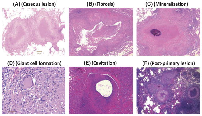
Different types of histopathological lesions observed during various Mtb infections of rhesus macaques. A. Centrally caseous lesion with peripheral rim of immune cells is the most typical type of pathology observed in NHPs infected with Mtb. B. A rare fibrotic lesion in a rhesus macaque infected with Mtb. C. Mineralization of a caseous lesion over time in a rhesus macaque infected with Mtb. D. Formation of highly inflamed multinucleated giant macrophages in a lesion from a rhesus macaque infected with Mtb. E. A rare lesion in a rhesus macaque infected with Mtb with a pathology that could be a precursor for cavitation. F. Post-primary lesions in the vicinity of primary, centrally caseous lesions in a rhesus macaque coinfected with Mtb and simian immunodeficiency virus.