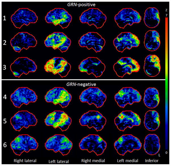
Fig. 1.
Stereotactic surface projection maps showing regions of hypometabolism in each GRN-positive and GRN-negative patient. All patients showed left parietal and temporal hypometabolism, which was particularly severe in patient 3, who also showed striking left frontal lobe hypometabolism. Less temporal lobe hypometabolism was observed in patient 6, who also showed a more bilateral pattern

