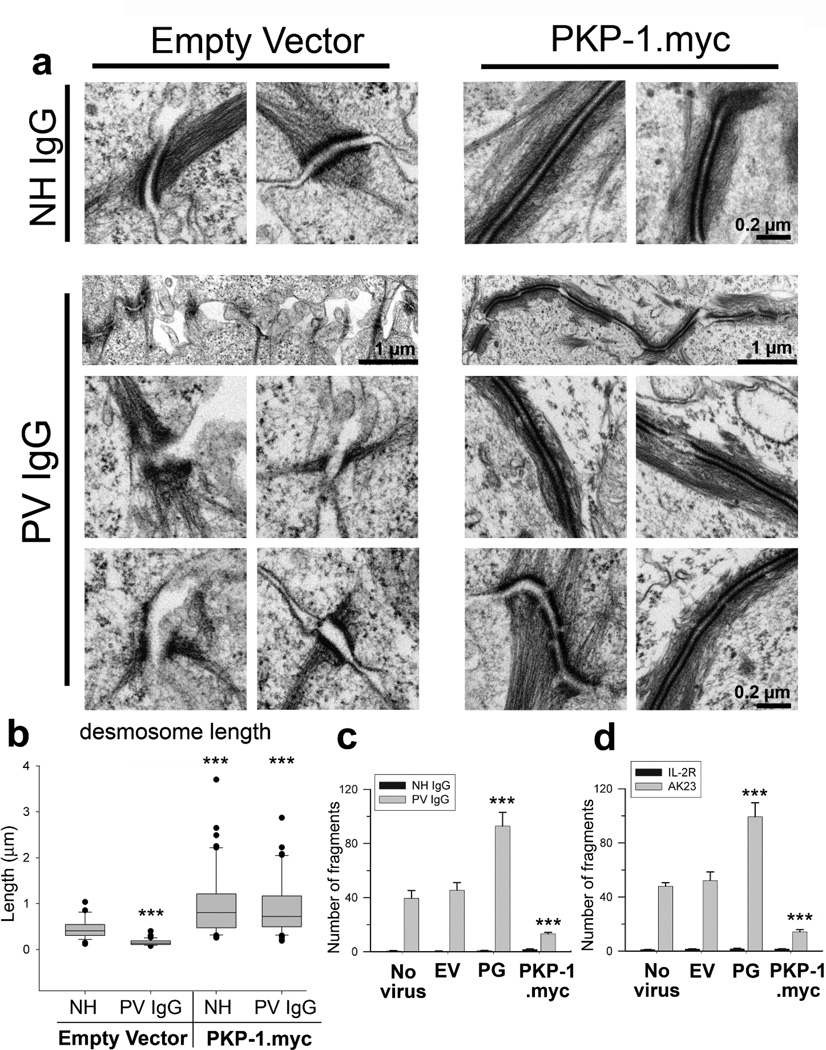
Figure 3.
PKP-1 protects desmosome ultrastructure and keratinocyte adhesion strength from disruption by PV IgG
(a) Electron micrographs and (b) quantification of desmosome lengths from keratinocytes expressing empty vector (EV) or PKP-1.myc treated with NH or PV IgG for 24 hours (n= 25–50 desmosomes per group); *** p < 0.001 compared with EV-NH IgG (Mann Whitney). (c, d) Quantification of monolayer fragmentation after cell dissociation assays using control keratinocytes (no virus, EV, and plakoglobin (PG)) or keratinocytes expressing PKP-1.myc exposed for 24 hours to (c) NH or PV IgG or (d) mAb IL-2R or AK23, a pathogenic Dsg3 mAb. Mean number of fragments ± SEM;*** p < 0.001 compared with no virus and EV (Two-way ANOVA, Holm-Sidak method). Scale bars, 0.2 or 1 Hm as indicated.

