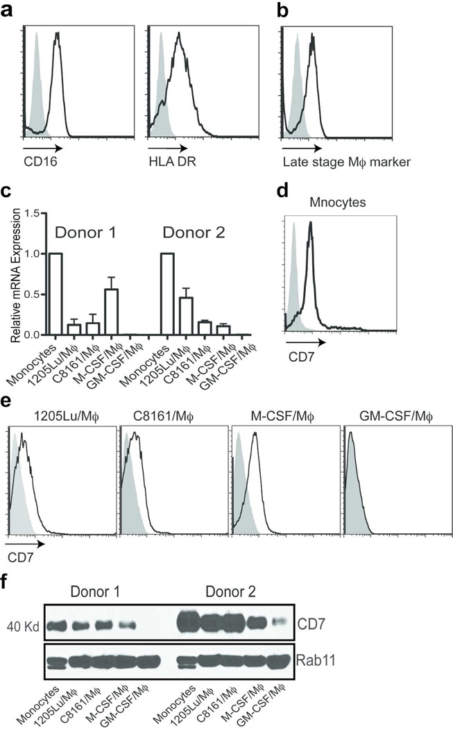
Figure 1. Expression of CD7 by monocytes and macrophages.
Expression of CD16, HLA DR (a) and the late stage maker (b) was analyzed by flow cytometry. 1205Lu-Mϕ) were stained with the fluorescence-conjugated anti-CD16, HLA DR and late stage macrophage marker. (c) Real-time PCR was used to analyze the expression of CD7 in monocytes, GM-CSF, M-CSF differentiated macrophages, and C8161 (C68161-Mϕ) and 1205Lu (1205Lu-Mϕ) melanoma conditioned media differentiated Mϕ. Samples were normalized to GAPDH. Monocytes (d) M-CSF/Mϕ, M-CSF/M(|), C8161/Mϕ) and 1205Lu/Mϕ) (e) were stained with the anti-human CD7 monoclonal antibody, 3A1, following by FITC-conjugated anti-mouse IgG staining for FACS analysis. Filled: Isotype control, black line: anti-CD7. (f) Western blot analysis expression of CD7 in monocytes, GM-CSF/Mϕ), M-CSF/Mϕ, and C8161/Mϕ and 1205Lu/Mϕ. RAb11 was used as a loading control.

