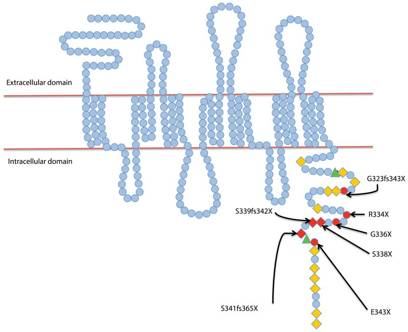
Figure 3. The structure of CXCR4.
Amino acids are represented as blue circles. The cytoplasmic tail is rich in serine (orange diamonds) and threonine (green triangles) residues. Sites of mutations are highlighted in red. The respective reported mutations involved in the WHIM syndrome are marked with arrows.

