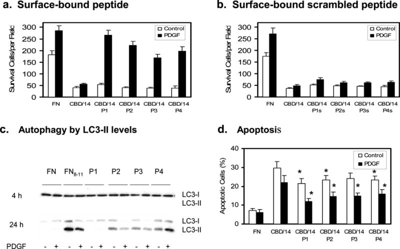
Figure 2. FN-GFB peptide coupled to CBD/14 supported FN-null fibroblast responsiveness to PDGF-BB.

Cells were plated in 96-well (a,b,d) or 6-well (c) plates precoated with 0.125 μM FN or glutathione S-transferase (GST)-tagged CBD/14±tethered peptide and cultured in serum-free DMEM±PDGF-BB at 37°C for 3d. a, b. Viable spread cells in five 10xfields were counted in three wells at 3d (mean ± SD, n = 15). c. LC3-II was detected by a size shift on Western blot using a polyclonal antibody specific for LC3 (Zhang et al 2007). d. Apoptosis was determined by TUNEL at 3d. Percent apoptosis was calculated from (positive-cells/total-cells)×100. Fifty cells were counted in 3 replicate plates. Data points indicate mean ± SD. Each panel represents at least 3 experiments. *p<0.05 compared with CBD/14.
