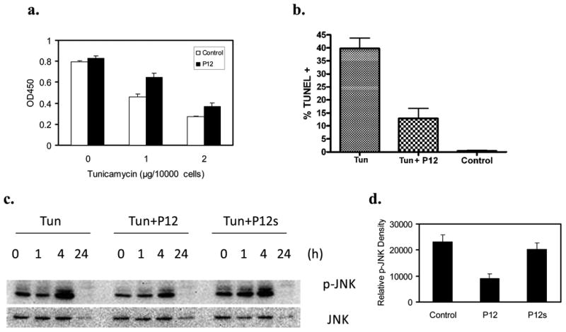
Figure 4. P12 enhanced AHDF survival under tunicamycin (Tun)-induced endoplasmic reticulum (ER) stress and suppressed Tun-induced JNK activation.

a. AHDF at 1000 cells/well were cultured in serum-free DMEM + 1nM PDGF-BB in 96-well plates overnight and challenged with Tun at 1 or 2μg/104cells for 4h. Cell viability was determined by XTT assay. Histograms show mean±SD, n=4. b. AHDF were incubated in serum-free DMEM + 1nM PDGF-BB with 1μg Tun/104cells±10μM P12 for 20h. Apoptosis was determined by TUNEL assay. c-d. AHDF were cultured in complete DMEM and exposed to 5μg/ml Tun (1μg/104cells)±10μM P12 or 10μM scrambled P12 (P12s) for the indicated times. c. p-JNK levels were determined by immunoblotting. d. Quantitative analysis of p-JNK band at 4h from 3 immunoblots, mean±SD.
