Figure 1.
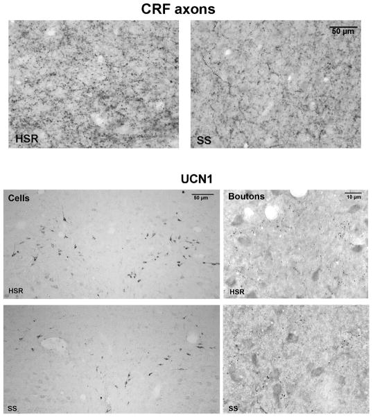
Figure 1, top. Photomicrographs of CRF axonal bouton staining in the dorsal raphe region of an HSR cynomolgus macaque obtained with a Leica brightfield microscope.
Left. CRF axonal bouton staining in an HSR animal.
Right. CRF axonal bouton staining in an SS animal.
Figure 1, bottom. Photomicrographs of UCN1 immunostained neurons and boutons at anatomically matching sections from representative HSR and SS monkeys obtained with a Leica brightfield microscope.
Left. The neurons are located in the supraocculomotor area (SOA) adjacent to the Edinger-Westfal nucleus, which is rostral to the raphe nucleus. There were more detectable neurons in the HSR monkey than the SS monkey, which was typical of the groups.
Right. The immunostained boutons are located caudal to the EWN and rostral to the raphe in a small consistent axonal cluster. There were similar numbers of UCN1 immunostained boutons in the HSR and SS groups, as observed in the representative animals shown here. Image subtraction was used in the bouton analysis and nonspecifically stained cells were removed to aid the threshold discrimination of boutons.

